Introduction to Implant Scan Body Selection and Scanning Protocol for Dentists in 2026
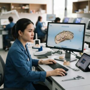
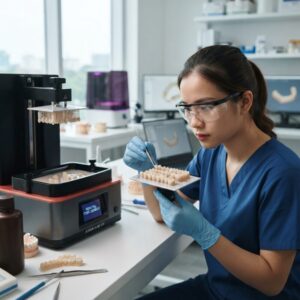
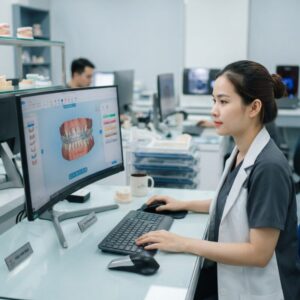
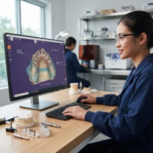
The implant scan body selection and scanning protocol for dentists in 2026 has evolved dramatically with the integration of cutting-edge CAD/CAM technology and intraoral scanning advancements. Proper selection and scanning protocols are foundational for achieving highly accurate digital impressions that translate flawlessly into implant-supported prosthetics. This article delves deeply into the nuances of selecting the right scan bodies and executing precise scanning protocols, tailored for dental labs, DSOs, and dental practices looking to optimize their implant workflows.
What Are Implant Scan Bodies and Why Are They Critical?
Implant scan bodies are specialized components that attach to dental implants during digital impression acquisition, serving as reference markers for implant position and orientation.
They are critical because they enable the intraoral scanner to capture the exact three-dimensional position of the implant, which is essential for designing accurate crowns, bridges, surgical guides, dentures, orthodontic aligners, and night guards. Without precise scan bodies, errors in prosthetic fit and function increase, leading to costly remakes and patient dissatisfaction.
Modern scan bodies are designed to be compatible with specific implant systems, featuring unique geometries and materials optimized for optical scanning. Their design directly impacts scan accuracy, ease of use, and the efficiency of digital workflows.
How Does the Implant Scan Body Selection and Scanning Protocol for Dentists in 2026 Improve Clinical Outcomes?
The implant scan body selection and scanning protocol for dentists in 2026 improves clinical outcomes by standardizing digital impression procedures to maximize accuracy and reduce chair time.
By choosing the appropriate scan body that matches the implant system and clinical scenario, and by following an optimized scanning protocol, clinicians can ensure that the digital data sent to the dental lab is precise and comprehensive. This leads to better fitting prosthetics, fewer adjustments, and enhanced patient comfort. Additionally, the protocol integrates seamlessly with CAD/CAM design software, supporting streamlined communication between dental offices and outsourcing labs.
What Are the Key Factors in Selecting Implant Scan Bodies in 2026?
Key factors in implant scan body selection include implant system compatibility, material properties, design features, and digital workflow integration.
- Implant System Compatibility: Scan bodies must precisely match the implant brand and model (e.g., Straumann, Nobel Biocare) to ensure accurate data capture.
- Material: Scan bodies made from PEEK, titanium, or high-grade plastics have different reflectivity and durability, impacting scan accuracy.
- Design: The geometry should promote easy seating, stable positioning, and minimal interference with surrounding soft tissues.
- Digital Workflow: Scan bodies should be supported by the intraoral scanner’s software and compatible with CAD platforms such as exocad or 3Shape.
Choosing the right scan body reduces errors and enhances the predictability of implant prosthetics, which is crucial for large DSOs managing multiple implant cases concurrently.
What Are the Standard Scanning Protocols for Implant Scan Bodies in 2026?
Standard scanning protocols for implant scan bodies in 2026 include a systematic approach to intraoral scanning that ensures comprehensive capture of implant position and surrounding anatomy.
The protocol typically involves:
- Pre-Scan Preparation: Confirm the scan body is properly seated and free of debris.
- Initial Scan: Capture the occlusal surfaces and adjacent teeth to establish landmarks.
- Focused Scan: Scan the scan body from multiple angles to capture full geometry.
- Soft Tissue Capture: Include surrounding gingiva to aid in accurate prosthetic design.
- Verification: Use scanning software to verify data completeness and quality in real-time.
Following these protocols minimizes the risk of rescans and improves the efficiency of the implant restorative process.
How Are New Technologies Shaping Implant Scan Body Selection and Scanning Protocols?
New technologies such as AI-enhanced scanning, improved intraoral scanners, and advanced CAD software are revolutionizing implant scan body selection and scanning protocols for dentists in 2026.
Artificial intelligence assists in automatically identifying scan bodies within the digital impression and optimizing scan paths to reduce operator error. Furthermore, the latest scanners from brands like 3Shape and Medit offer higher resolution and faster data capture, facilitating smoother workflows.
CAD software platforms now provide direct integration with implant manufacturer libraries, streamlining the design process and enabling outsourcing labs to deliver prosthetics with superior fit and esthetics. These advancements contribute significantly to reducing turnaround times and improving clinical predictability.
What Are the Common Challenges in Implant Scan Body Selection and How to Overcome Them?
Common challenges include incorrect scan body choice, improper seating, scanning artifacts, and software incompatibility.
Incorrect selection or mixing of scan bodies from different implant systems leads to inaccurate data and ill-fitting prosthetics. Clinicians should verify implant system details before ordering scan bodies. Proper seating involves tactile confirmation and visual inspection to ensure full engagement without movement.
Scanning artifacts can arise from reflective surfaces or limited access in posterior regions; using scan bodies with matte finishes and employing strategic scanning angles can mitigate this. Ensuring that the intraoral scanner software supports the chosen scan body library is essential for seamless data processing.
How Does Outsourcing CAD/CAM Design Benefit from a Defined Implant Scan Body Selection and Scanning Protocol?
Outsourcing CAD/CAM design benefits greatly from a defined implant scan body selection and scanning protocol for dentists in 2026 by guaranteeing the quality and consistency of digital impressions submitted to dental labs.
When dental practices adhere to a standardized protocol, outsourcing labs can reliably process digital files, reducing the need for back-and-forth communications and remakes. This efficiency translates into faster turnaround times and cost savings for DSOs managing multiple locations. Furthermore, labs can integrate the digital data directly into implant prosthetic design software, such as exocad or 3Shape, enhancing precision and workflow automation.
What Role Do Surgical Guides Play in Implant Scan Body Scanning Protocols?
Surgical guides are critical adjuncts in implant placement planning and benefit from accurate implant scan body scanning protocols.
Accurate scanning of implant scan bodies ensures precise digital impressions that guide the fabrication of surgical guides, enabling controlled and predictable implant placement. Digital impressions that capture the implant position and surrounding anatomy allow dental labs to design guides that fit perfectly and reduce intraoperative errors. This process also works well alongside surgical guide design services, enhancing overall implant treatment success.
What Are Best Practices for Dentists When Implementing the Implant Scan Body Selection and Scanning Protocol for Dentists in 2026?
Best practices include continuous training, investing in compatible scanning technology, and collaborating closely with dental labs.
- Training: Regular staff training on scan body handling and scanning techniques to maintain high data quality.
- Technology: Using updated intraoral scanners with comprehensive implant libraries from trusted manufacturers.
- Communication: Establishing clear communication channels between the dental office and outsourcing lab to clarify preferences and resolve issues rapidly.
- Quality Checks: Implementing routine verification of scan data prior to submission to minimize errors.
Adhering to these best practices not only optimizes the implant scan body selection and scanning protocol for dentists in 2026 but also enhances patient outcomes and practice efficiency.

How Are Digital Dentures and Orthodontic Aligners Impacted by Implant Scan Body Protocols?
Digital dentures and orthodontic aligners increasingly rely on precise implant scan body protocols to ensure seamless integration with implant-supported prosthetics.
For implant-retained dentures, accurate digital impressions of scan bodies enable the correct positioning of attachments and frameworks. Similarly, orthodontic aligners used in complex cases involving implants require accurate scanning to avoid misfits and ensure effective tooth movement. The implant scan body selection and scanning protocol for dentists in 2026 supports these workflows by providing reliable digital data that can be integrated with denture design and orthodontic software platforms.
What External Resources Are Recommended for Staying Updated on Implant Scan Body Technologies?
Leading dental organizations and implant manufacturers provide invaluable resources for staying current on implant scan body technologies and scanning protocols.
Websites such as the American Dental Association (ADA) offer clinical guidelines and continuing education. Implant system manufacturers like Straumann and Nobel Biocare provide detailed product information and digital workflow recommendations. Additionally, software providers like exocad publish updates on implant libraries and scanning standards.
Leveraging these resources helps dental professionals and labs maintain proficiency with evolving implant scan body selection and scanning protocol for dentists in 2026.
Conclusion: Why Prioritizing Implant Scan Body Selection and Scanning Protocol for Dentists in 2026 Matters
Prioritizing the implant scan body selection and scanning protocol for dentists in 2026 is essential for delivering high-quality implant prosthetics with efficiency and predictability. This protocol ensures that digital impressions are accurate, reproducible, and fully compatible with modern CAD/CAM design software and outsourcing workflows.
As dental labs and DSOs increasingly adopt digital solutions, a well-defined protocol minimizes errors, reduces remakes, and accelerates case turnaround times, ultimately benefiting both clinicians and patients.
Contact Us
Step-by-Step Guide to Implementing an Effective Implant Scan Body Workflow
Introducing a robust workflow for implant scan body selection and scanning protocol can significantly elevate the accuracy and efficiency of implant restorations. Below is a detailed step-by-step guide designed for dental practices and labs to streamline this process and minimize errors.
Step 1: Pre-Scan Preparation and Case Assessment
- Review Patient Records: Begin by thoroughly reviewing the patient’s medical and dental history, radiographs, and any previous digital scans.
- Identify Implant System: Confirm the exact implant brand, model, and platform dimensions. This is crucial because scan bodies are specific to implant systems.
- Choose Appropriate Scan Body: Select a scan body compatible with the implant. Consider material (PEEK, titanium), design (one-piece or two-piece), and manufacturer recommendations.
- Prepare the Scan Body: Clean and sterilize the scan body according to the manufacturer’s protocols. Verify that the scan body is free of defects or damage.
Step 2: Scan Body Placement and Verification
- Securely Attach Scan Body: Attach the scan body firmly to the implant analog or directly intraorally, ensuring it is seated fully without gaps.
- Visual Inspection: Use magnification to confirm correct orientation and seating. Misplacement can cause digital distortion.
- Dry the Area: Ensure the implant site and scan body are dry to reduce scan reflection and artifacts.
Step 3: Scanning Protocol Execution
- Calibrate Scanner: Perform scanner calibration per the manufacturer’s guidelines to ensure accuracy.
- Scan Surrounding Anatomy First: Capture adjacent teeth and soft tissue landmarks before scanning the scan body for better spatial reference.
- Scan Scan Body: Capture multiple angles of the scan body, moving slowly to avoid stitching errors or data loss.
- Confirm Scan Completeness: Review the digital impression for missing data, voids, or distortions, especially around the scan body.
Step 4: Data Transfer and CAD Integration
- Export Scan Files: Export STL or proprietary scan files with clear labeling for implant site and scan body used.
- Communicate with Lab: Provide detailed instructions to the dental lab or CAD/CAM technician, including implant details and any special considerations.
- Verify CAD Design: Upon receiving the CAD design, verify that the digital model aligns perfectly with the scan body data.
Step 5: Final Verification and Adjustment
- Try-in Prosthetic: Once the prosthesis is fabricated, perform a clinical try-in to verify fit and occlusion.
- Make Necessary Adjustments: If discrepancies occur, identify whether they stem from scan inaccuracies or design errors.
- Implement Feedback Loop: Document issues and share feedback with the team or lab to improve future scan protocols.
Common Mistakes in Implant Scan Body Selection and Scanning—and How to Avoid Them
Despite advances in technology, common pitfalls undermine the success of implant scanning workflows. Understanding and proactively addressing these mistakes can prevent costly remakes and delays.
1. Using Non-Compatible or Generic Scan Bodies
Many practitioners opt for generic or universal scan bodies to save costs. However, these often lack precise fit and may not capture the implant interface accurately, leading to ill-fitting restorations.
- Tip: Always verify implant compatibility and use manufacturer-approved scan bodies to ensure precision.
2. Incorrect Scan Body Seating
Improper attachment—such as partial seating, cross-threading, or loose fixation—results in inaccurate scan data and compensatory errors in the final prosthesis.
- Tip: Use torque drivers or manufacturer-recommended tools when securing scan bodies. Confirm seating visually and tactilely.
3. Inadequate Scanner Calibration and Maintenance
Failing to regularly calibrate or maintain intraoral scanners can degrade scan accuracy, introduce noise, and increase scanning time.
- Tip: Integrate routine scanner maintenance schedules and conduct pre-scan calibration checks.
4. Insufficient Scanning Angles and Coverage
Scanning the scan body from a single angle or too quickly may lead to incomplete data capture and stitching errors in the software.
- Tip: Capture multiple angles and slow, deliberate scanning passes around the scan body.
5. Ignoring Soft Tissue and Surrounding Anatomy
Not scanning adjacent teeth and soft tissue landmarks can cause spatial inaccuracies when aligning the implant position digitally.
- Tip: Always include at least the adjacent teeth and gingival margins to provide a full reference framework.
6. Poor Communication Between Clinic and Lab
Unclear or incomplete case instructions regarding implant details, scan body types, or patient-specific nuances can result in design errors and remakes.
- Tip: Use standardized digital work orders and include photos or videos if necessary to clarify instructions.
Cost and Quality Considerations When Choosing Implant Scan Bodies
Balancing cost and quality is a critical concern for dental labs and practices adopting implant scanning protocols. Here’s how to approach these factors pragmatically.
Material Impact on Cost and Accuracy
Scan bodies are commonly made from materials like PEEK (polyether ether ketone), titanium, or composite resins. Each has specific cost and performance profiles:
- PEEK: Lightweight, radiolucent, and cost-effective. Provides good scan data quality but may wear faster with repeated use.
- Titanium: Highly durable and precise but more expensive. May cause reflection artifacts in scanning if not coated properly.
- Composite Resin: Mid-range cost, with varying accuracy based on manufacturer quality.
Reusable vs. Single-Use Scan Bodies
Reusable scan bodies reduce long-term costs but require strict sterilization protocols and risk wear. Single-use scan bodies eliminate cross-contamination risk but increase per-case expenses.
- Recommendation: Practices with high implant volume may benefit from investing in high-quality reusable scan bodies paired with strict sterilization procedures.
Quality Assurance Through Standardization
Implementing standardized protocols for scan body handling, scanner calibration, and data transfer minimizes costly errors and remakes.
- Invest in staff training to maintain consistent scan quality.
- Use quality control checklists to verify each step in the scanning workflow.
- Monitor and document restoration fit outcomes to identify trends and areas for improvement.
Budgeting for Technology Investments
Adopting advanced scanning systems and high-quality scan bodies requires upfront investment but reduces long-term costs associated with remakes and chairside adjustments.
- Consider total cost of ownership, including software updates, service contracts, and consumables.
- Evaluate return on investment by tracking case turnaround times and patient satisfaction.
Market Specificities: Adapting Implant Scan Body Selection and Scanning Protocols to Regional and Practice Variations
Market conditions, regulatory environments, and patient demographics can influence implant workflows and scan body selection. Tailoring protocols to these specifics enhances clinical success and business viability.
Regional Brand Availability and Preferences
Implant systems and scan body manufacturers vary significantly by region due to distribution agreements, regulatory approvals, and market preferences. For example:
- Europe: Strong presence of certain implant brands like Straumann and Astra Tech may dictate scan body selection.
- Asia-Pacific: Growing adoption of emerging manufacturers with cost-competitive scan bodies.
- North America: Preference for well-established brands with extensive digital workflows and third-party scan body options.
Dental practices should maintain an updated inventory of compatible scan bodies aligned with their implant portfolio and local market availability.
Practice Size and Workflow Complexity
Small private practices and large DSOs differ in implant volume, staffing, and technology budgets, requiring customized approaches:
- Small Practices: May benefit from simplified protocols and partnerships with local labs to manage costs.
- Large DSOs: Can implement centralized scanning centers with standardized protocols, enabling bulk purchasing and dedicated digital specialists.
Patient Demographics and Anatomical Considerations
Population-specific factors such as bone density, oral anatomy, and prevalence of certain implant types influence scan body choice and scanning strategy. For example:
- Elderly patients with compromised bone quality may require scan bodies that offer enhanced scanning contrast.
- Patients with limited mouth opening necessitate smaller scan bodies or modified scanning techniques.
Regulatory and Reimbursement Factors
Understanding local regulatory guidelines regarding device use and reimbursement policies for digital workflows can impact protocol adoption.
- Some regions may require traceability documentation for scan bodies and digital impressions as part of quality control.
- Inclusion of digital scanning in insurance reimbursement can incentivize investment in advanced protocols.
Adapting to Emerging Market Trends
Keeping abreast of evolving market trends such as the rise of tele-dentistry, remote design collaboration, and AI-assisted scanning can provide competitive advantages.
- Practices in emerging markets may leapfrog traditional workflows by adopting cloud-based scanning and design solutions.
- Flexible implant scan body protocols that integrate with these innovations will support scalability and enhanced patient care.
Ready to enhance your implant workflows with expert CAD/CAM outsourcing? Contact us today to learn how our digital design services can support your practice’s success.