Introduction to How to Evaluate Crown Fit and Contacts Before Cementation in 2026
Understanding how to evaluate crown fit and contacts before cementation in 2026 is essential for dental labs and DSOs aiming to deliver high-quality restorations. As digital workflows and CAD/CAM technology evolve, the precision and predictability of crown fabrication have improved significantly. However, ensuring that each crown fits perfectly and has appropriate occlusal and proximal contacts remains a critical step before cementation to avoid clinical failures such as marginal gaps, patient discomfort, or secondary caries.
This comprehensive guide explores state-of-the-art evaluation methods, combining digital tools with proven clinical techniques. It is designed to help dental professionals and lab technicians optimize their crown assessment processes, reduce remakes, and enhance patient outcomes.
Why Is Precise Evaluation of Crown Fit and Contacts Crucial in Modern Dentistry?
Precise evaluation of crown fit and contacts is vital to preserve tooth structure, maintain periodontal health, and ensure long-term restoration success. Poor crown fit can lead to microleakage, cement washout, and eventual restoration failure, while improper contacts may cause food impaction or occlusal trauma.
In 2026, the integration of digital design and manufacturing technologies demands that dental labs and clinics adopt rigorous verification protocols. These protocols minimize human error and enhance the accuracy of fit and contacts through a combination of digital analysis and clinical simulation. This is especially important given the rise of complex restorations like implant crowns, surgical guides, and orthodontic appliances, which require precise adaptation.
What Are the Latest Digital Tools for Evaluating Crown Fit and Contacts?
The answer: Leading-edge CAD/CAM software and intraoral scanners now provide highly accurate 3D models to evaluate crown fit and contacts digitally before physical fabrication or clinical try-in.
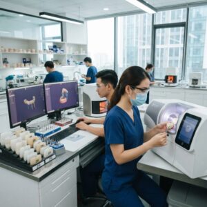
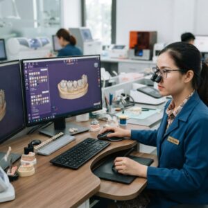
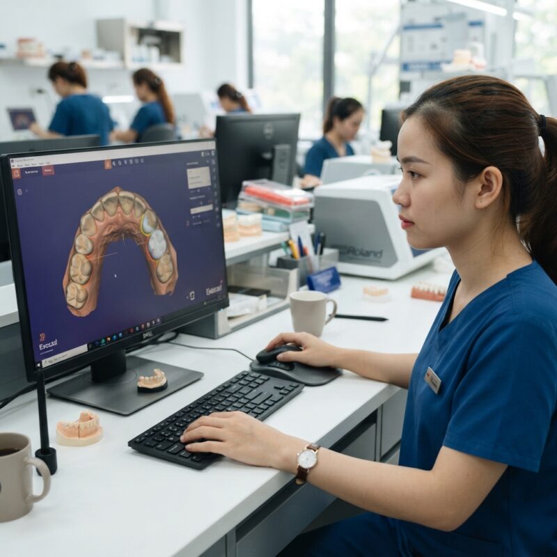
Modern systems from providers such as 3Shape and exocad offer advanced tools for visualizing marginal gaps and contact points. These platforms enable dental technicians to simulate occlusion, check proximal contacts, and adjust restorations virtually. For example, digital occlusal analysis can highlight high spots or undercontacts using color-coded maps, allowing for precise modifications before milling or printing. This reduces chairside adjustments and improves patient comfort.
Moreover, intraoral scanners like those from iTero facilitate real-time assessment of crown fit by capturing detailed 3D impressions directly from the patient. This data can be cross-referenced with the CAD design to detect discrepancies early.
What Are the Traditional Clinical Techniques for Checking Crown Fit and Contacts?
Traditional clinical techniques remain indispensable even in the digital era for confirming crown fit and contacts before cementation. The direct answer is that tactile and visual methods such as using fit-checking materials, articulating paper, and radiographs are still gold standards.
Fit-checking materials like silicone or pressure-indicating paste allow clinicians to detect internal gaps or pressure points by seating the crown on the prepared tooth and observing the material thickness. Articulating paper is used to identify occlusal contact points and ensure even distribution of forces. Proximal contacts are tested with dental floss or shimstock to confirm that contacts are neither too tight nor too loose, preventing food impaction or patient discomfort.
Additionally, periapical or bitewing radiographs can verify marginal adaptation and confirm absence of overhangs or voids that may compromise cementation. These methods complement digital evaluations and provide a comprehensive assessment protocol.
How to Evaluate Crown Fit and Contacts Before Cementation in 2026 Using a Hybrid Workflow?
Combining digital and clinical techniques into a hybrid workflow is the most effective approach for how to evaluate crown fit and contacts before cementation in 2026. This approach leverages the precision of CAD/CAM design with the reliability of traditional clinical verification.
- Step 1: Digital Pre-Evaluation – Use CAD software to analyze crown design, focusing on margin contours and contact points. Simulate occlusal contacts with digital articulation tools.
- Step 2: Fabrication – Manufacture the crown with high-precision milling or 3D printing from verified digital files.
- Step 3: Clinical Try-In – Conduct a physical assessment of crown fit using pressure-indicating materials and articulating paper.
- Step 4: Contact Adjustment – Adjust proximal contacts with finishing strips and occlusal contacts with fine burs or polishers, guided by clinical feedback.
- Step 5: Radiographic Confirmation – Take bitewing or periapical radiographs to confirm margin adaptation and contact integrity.
This hybrid method minimizes remakes and ensures that the crown will function optimally once cemented. It also works well alongside surgical guide design services, which require similar precision.
What Role Does Dental Lab Outsourcing Play in Crown Fit Evaluation?
Dental lab outsourcing plays a significant role by providing expert CAD/CAM digital design and fit verification services that many dental clinics and DSOs may not have in-house. The direct answer is that outsourcing to specialized labs streamlines the evaluation process and elevates crown quality through advanced technology and experienced technicians.
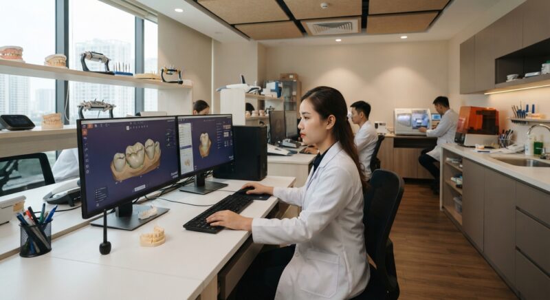
Outsourced labs use cutting-edge software and scanning devices to analyze crown fit and contacts thoroughly before sending restorations to the clinic. They can simulate various clinical scenarios and optimize crown design for ideal margin adaptation and contact tightness. This reduces chairside adjustments and enhances patient satisfaction.
Partnering with an outsourcing lab also provides access to a broader range of expertise for complex cases, such as implant crowns or full-arch prostheses, which benefit from meticulous fit evaluation. Leading labs often integrate with intraoral scanning workflows from platforms like Medit, ensuring seamless digital communication and quality control.
What Are Common Challenges in Evaluating Crown Fit and Contacts and How to Overcome Them?
Even with advanced techniques, challenges persist in how to evaluate crown fit and contacts before cementation in 2026. Common issues include inaccurate digital impressions, material shrinkage during fabrication, and subjective clinical assessments.
To overcome these challenges, the following strategies are recommended:
- Use High-Quality Scanners: Ensure intraoral scans capture full margin details and proximal areas accurately.
- Calibrate Milling/Printing Equipment: Regular maintenance and calibration reduce errors from material distortion.
- Standardize Clinical Protocols: Train clinicians on consistent use of fit-checking materials and contact verification tools.
- Leverage Software Validation: Utilize CAD software that offers automated gap analysis and contact strength indicators.
- Implement Feedback Loops: Establish communication channels between labs and clinics for continuous improvement based on clinical outcomes.
Adhering to these best practices ensures that both digital and clinical methods align for precise crown evaluation.
How Does Material Selection Affect the Evaluation Process?
The choice of restorative material significantly influences how to evaluate crown fit and contacts before cementation in 2026. Different materials have varying properties such as shrinkage, flexibility, and translucency that impact fit and contact accuracy.
For example, zirconia crowns require sintering, which can cause dimensional changes that must be anticipated during design and evaluation. Lithium disilicate crowns, often milled fully crystallized, offer excellent fit but may require careful occlusal adjustment due to brittleness. PMMA or resin-based materials used for provisional crowns allow easier intraoral modification but demand verification of marginal seal before cementation.
Understanding these material characteristics helps technicians and clinicians tailor their evaluation protocols. Digital software often includes material-specific parameters that predict fit changes during processing, aiding in preemptive design modifications.
What Are the Best Practices for Documenting Crown Fit and Contact Evaluations?
Documenting the evaluation process is crucial for quality assurance, traceability, and continuous improvement. The direct answer is that comprehensive records including digital design files, clinical photographs, and evaluation notes ensure accountability and facilitate communication between labs and clinicians.
Best practices include:
- Saving digital scans and CAD designs with annotations of fit and contact adjustments.
- Capturing intraoral photographs of trial seating showing fit-check material distribution.
- Recording articulating paper markings and floss test results for proximal contacts.
- Maintaining radiographic documentation confirming margin integrity.
- Using digital platforms that allow secure sharing of evaluation data with stakeholders.
Robust documentation supports the reduction of remakes and enhances patient care by allowing retrospective analysis of any issues that arise.
Conclusion and Call to Action
Mastering how to evaluate crown fit and contacts before cementation in 2026 requires embracing both cutting-edge digital tools and time-tested clinical techniques. By adopting a hybrid workflow, utilizing expert outsourcing services, and adhering to best practices in material handling and documentation, dental labs and DSOs can achieve superior restoration outcomes.
Step-by-Step Checklist for Crown Fit and Contact Evaluation Before Cementation
Ensuring a successful crown fit and optimal contacts requires a systematic approach. The following checklist provides a practical, step-by-step guide dental professionals and lab technicians can follow before cementation:
- Step 1: Initial Visual Inspection
- Examine the crown under magnification for obvious defects such as voids, sharp margins, or overcontoured areas.
- Verify shade and translucency match with adjacent teeth to avoid esthetic discrepancies.
- Step 2: Try-in and Seating Check
- Gently seat the crown on the prepared tooth without cement to check passive fit.
- Use articulation paper to detect any premature occlusal contacts.
- Check for rocking or gaps by applying gentle finger pressure and slight lateral forces.
- Step 3: Marginal Gap Measurement
- Employ magnification tools or dental loupes to inspect crown margins closely.
- Use fit-checking materials (e.g., disclosing wax or silicone fit indicators) to confirm margin adaptation.
- Note any discrepancies and communicate necessary adjustments to the lab if gaps exceed clinical thresholds (commonly >120 microns).
- Step 4: Proximal Contact Assessment
- Use dental floss or thin strips to test contact tightness on both mesial and distal sides.
- Ensure contacts are not too tight (which may cause patient discomfort or food impaction) or too loose (leading to drifting or plaque accumulation).
- Adjust proximal contours as needed using fine burs or by requesting lab modification.
- Step 5: Occlusal Contact Verification
- Use articulating paper in multiple jaw positions (centric occlusion, protrusive, lateral excursions) to identify high spots.
- Mark and adjust occlusal contacts to distribute forces evenly and avoid premature contacts that can lead to crown fracture or patient discomfort.
- Step 6: Final Documentation
- Record findings including photographs, notes on adjustments, and any communication with the dental lab.
- Use digital records integrated into practice management software to streamline follow-up and quality control.
Common Mistakes in Crown Fit and Contact Evaluation and How to Avoid Them
Even experienced clinicians and technicians can encounter pitfalls during crown fit assessments. Awareness of these common mistakes can prevent costly remakes and patient dissatisfaction.
- Relying Solely on Visual Inspection Without Quantitative Measurement
Visual checks are subjective and can miss marginal gaps or subtle contact issues. Incorporating fit-checking materials, digital scans, or microscopes enhances accuracy.
- Ignoring Patient Feedback During Try-In
Patients may notice discomfort from tight contacts or occlusal discrepancies before the clinician. Always solicit and address patient input before cementation.
- Failing to Check Contacts in Multiple Jaw Positions
Occlusal contacts can vary in dynamic movements. Checking only centric occlusion risks overlooking problematic contacts during lateral or protrusive excursions.
- Over-Adjusting Proximal Contacts Chairside
Excessive grinding can weaken the crown or remove critical contours. Minor adjustments are acceptable, but significant changes should be referred back to the lab for recasting or redesign.
- Not Accounting for Material-Specific Properties
Different crown materials behave differently during adjustment and cementation. For example, zirconia crowns can chip if aggressively ground, while metal crowns are more forgiving.
- Skipping Documentation and Communication With the Lab
Failure to document fit issues and communicate with the dental lab can lead to repeated errors and delays. Clear feedback loops improve future crown fabrication accuracy.
Cost vs. Quality Considerations in Crown Fit Evaluation Workflows
Balancing cost-effectiveness with high-quality crown fit evaluations is a strategic concern for both dental practices and labs. The following factors can influence this balance:
Investing in Digital Technologies
Advanced intraoral scanners, digital microscopes, and 3D fit analysis software can significantly improve evaluation precision but come with upfront costs. Practices and labs should consider:
- Return on investment (ROI) through reduced remakes and chairside adjustment times.
- Training costs and learning curves for staff.
- Integration with existing practice management and lab workflows.
Laboratory Outsourcing vs. In-House Evaluation
Outsourcing fit evaluation to specialized labs can save costs on equipment and expertise but may introduce delays and reduce direct control. In-house evaluation empowers immediate adjustments but requires investment in technology and training.
Material Costs and Impact on Evaluation
Some materials demand more precise fit due to their brittleness or esthetic sensitivity. For instance, all-ceramic crowns may require stricter margin adaptation, increasing evaluation time and potential costs.
Cost-Saving Tips Without Compromising Quality
- Implement standardized checklists and protocols to streamline evaluation and reduce unnecessary steps.
- Train dental assistants or hygienists to perform preliminary fit checks, freeing clinicians for complex assessments.
- Use cost-effective fit-checking materials like silicone indicators and articulating papers judiciously.
- Foster open communication with labs to minimize remakes and adjustments through clear prescriptions and feedback.
Market-Specific Considerations: Tailoring Crown Fit Evaluations to Different Dental Practice Models
The approach to crown fit and contact evaluation can differ widely depending on the type of dental practice or lab. Understanding these market-specific nuances helps optimize workflows and patient outcomes.
Private Practices vs. Dental Service Organizations (DSOs)
- Private Practices:
Usually have more personalized workflows and direct communication with labs. They benefit from flexible evaluation protocols tailored to individual patient needs but may have limited access to cutting-edge technology due to budget constraints.
- DSOs:
Often manage high patient volumes and multiple clinics, necessitating standardized evaluation protocols and digital integration for consistency. DSOs may invest more heavily in technology to maximize efficiency but face challenges coordinating across locations.
Dental Laboratories: Commercial vs. In-House Labs
- Commercial Labs:
Handle a high volume of cases with diverse materials and techniques. They rely on standardized digital and analog evaluation protocols to ensure consistent quality. Communication with multiple clinics requires robust documentation and digital data exchange.
- In-House Labs:
Embedded within dental practices, these labs facilitate rapid feedback and adjustments. They can offer more customized evaluations but may be limited in technology compared to large commercial labs.
Regional and Regulatory Considerations
Different regions may have varying regulations or standards regarding crown fit tolerances, materials used, and evaluation protocols. Staying abreast of these ensures compliance and enhances patient safety.
Adapting to Patient Demographics and Expectations
Practices serving diverse populations may need to adjust evaluation criteria based on factors such as occlusal habits, aesthetic demands, or economic considerations. For example, pediatric or geriatric patients might require more conservative proximal contact adjustments to accommodate sensitivities.
Contact us today to learn how our advanced CAD/CAM digital design outsourcing services can help you streamline crown fit evaluation and deliver exceptional results for your patients.